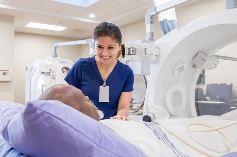
Radiographic procedures refer to medical imaging techniques used to visualize the internal structures of the body for diagnostic purposes. These procedures primarily utilize X-rays, a form of electromagnetic radiation, to produce images of bones, organs, and tissues. Radiographic procedures are performed to diagnose and monitor various medical conditions, guide surgical procedures, and assess the effectiveness of treatments. While X-rays involve exposure to ionizing radiation, modern radiographic techniques are designed to minimize radiation doses to patients, using protective measures and advanced equipment. Patients may need to remove jewelry and wear a hospital gown to avoid interference with the imaging process. Specific preparations might be required depending on the type of radiographic procedure. During the procedure, the patient is positioned appropriately, and the X-ray machine is adjusted to target the specific area of interest. The patient must remain still to ensure clear images are captured.

Radiographic Procedures
Radiologists are specialized medical doctors who have extensive training in interpreting medical images produced by radiographic procedures. Their expertise is crucial in diagnosing and managing a wide range of health conditions. X-rays pass through the body and are absorbed in varying degrees by different tissues. Dense tissues, like bones, absorb more X-rays and appear white on the radiograph, while softer tissues allow more X-rays to pass through and appear darker.